Institute Colloquium: Genetics and cell biology of complex cell shapes
Date
Monday, April 30, 2012 16:30 - 17:30
Speaker
Maria Leptin (EMBO)
Location
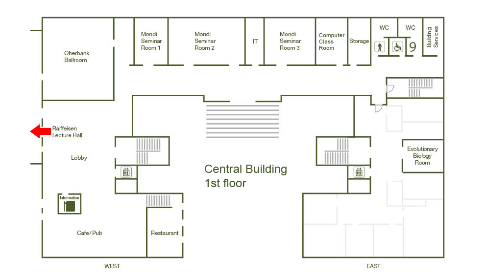
Raiffeisen Lecture Hall, Central Building
Series
Colloquium
Tags
Institute Colloquium
Contact

The terminal cells of the Drosophila respiratory (tracheal) network contain seamless, membrane bounded intracellular tubes through which air travels. The tube-containing extensions of these cells are elaborated during chemotactic growth directed by developmental cues in embryos and subsequently ramify in response to signals from hypoxic tissues in larvae. Extensive membrane traffic occurs during growth, as the cell rapidly and concomitantly elaborates two membrane domains of opposite characteristics. The outer (basal) membrane migrates and grows with highly dynamic filopodial extensions, while at the same time the inner tube of apical characteristics is constructed from as yet unknown membrane sources. We find that the membranes forming the intracellular tubules contain lipids and proteins typical of apical plasma membranes in polarized epithelial cells. The Drosophila synaptotagmin-like protein Bitesize (Btsz) and the activated form of its interaction partner Moesin are also located at the growing luminal membrane. Our functional studies indicate that the actin cytoskeleton, through its interaction with Btsz via Moesin directs apical membrane morphogenesis to create and maintain distinct intracellular tubules. Using real-time in vivo imaging we have analysed the assembly of the intracellular tube. We find that ER and Golgi rapidly distribute into the developing cellular extensions prior to the assembly of the membrane bounded tube within. Redistribution of secretory machinery ahead of the growing tube seems to be a prerequisite for proper tube extension.