Institute Colloquium: In vivo Connectivity: Paramagnetic Tracers, Electrical Stimulat
Date
Monday, April 16, 2012 16:30 - 17:30
Speaker
Nikos K. Logothetis (Max Planck Institute for Biological Cybernetics)
Location
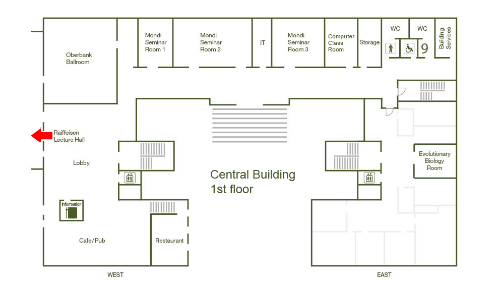
Raiffeisen Lecture Hall, Central Building
Series
Colloquium
Tags
Institute Colloquium
Contact

Neuroanatomical cortico-cortical and cortico-subcortical connections have been examined mainly by means of degeneration methods and anterograde and retrograde tracer techniques. Although such studies have demonstrated the value of the information gained from the investigation of the topographic connections between different brain areas, they do require fixed, processed tissue for data analysis and therefore cannot be applied to animals participating in longitudinal studies. Capacities such as plasticity and learning are indeed best studied with non-destructive techniques that can be applied repeatedly and, ideally, combined with neuroimaging or electrophysiology studies. The recent development of MR-visible tracers that can be infused into a specific brain region and are transported anterogradely transsynaptically is one such technique. Simultaneous electrical stimulation (ES) and fMRI (esfMRI) is another. In fact, esfMRI offers a unique opportunity for visualizing the networks underlying electrostimulation-induced behaviors, to map the neuromodulatory systems, or to study the effects of regional synaptic plasticity, e.g. LTP in hippocampus, on cortical connectivity. In my talk Ill present new data on MR-visible tracers and esfMRI that show the capacity of these methods for the study of the organization of cortical microcircuits and effective connectivity. I shall also show first results from studies mapping network topologies by triggering imaging at structure-specific events, e.g. hippocampal ripples or cross-frequency coupling events.