The Institute Colloquium: Two-photon optogenetics
Date
Monday, January 30, 2012 16:30 - 17:30
Speaker
Valentina Emiliani (Paris Descartes University)
Location
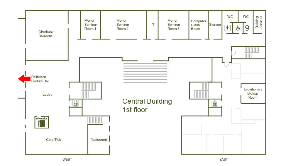
Raiffeisen Lecture Hall, Central Building
Series
Colloquium
Tags
Institute Colloquium
Contact

The combination of light microscopy and optogenetics actuators and reporters offers the possibility to control activation and inhibition of neuronal activity and monitor functional responses in a non-invasive manner enabling the analysis of well-defined neuronal population within intact neuronal circuits and systems. Interestingly, those tools have permitted to address key biological questions with relatively simple illumination methods using widefield visible light illumination. However, some limitations in the specificity of genetic targeting and the intricate morphology of the brain (neuronal processes, such as dendrites and axons, can reach regions far away from the cell soma), make it challenging to, for example, individuate subsets of genetically identical interconnected cells, or to establish the role of specific spatiotemporal excitatory patterns in guiding animal behavior. To reach such degree of specificity, more sophisticated illumination methods are required, permitting control of light patterning deep inside tissues. Here we present a novel microscopy system for high-resolution patterned photoactivation of optogenetics molecules based on the temporal control of ultrafast pulses for axial localization of the illumination pattern. For lateral light patterning, the system can use either digital holography [1-3] or the generalized phase contrast method [4]. We apply sculpted two-photon illumination to activate Channelrhodopsin-2 in mouse cultured neurons and cortical slices with sufficient efficacy to reliably fire action potentials with millisecond temporal resolution and low excitation power when the light was shaped over the cell body, one or more dendritic subdomains or multiple cells simultaneously [4].